The Mass Spectrometry Imaging Facility aims to provide high-quality, spatially resolved biomolecular data from tissue sections to support cancer research. Using Matrix Assisted Laser Desorption/Ionization (MALDI) across three complementary mass spectrometry platforms, the facility detects proteins, peptides, lipids, and metabolites. The Facility is supported by a Cancer Prevention and Research Institute of Texas (CPRIT) grant.

Mass Spectrometry Imaging (MSI) involves collecting thin tissue sections onto mass spectrometer-compatible slides. After potential sample preparation (washing, fixation, enzymatic digestion, matrix application), spectra are collected in an ordered array, resulting in images that show the spatial localization and intensity of biomolecules.
The facility focuses on MALDI for ion generation, which involves applying a chemical matrix to the tissue surface. A pulsed laser desorbs and ionizes molecules for detection. The MSI facility offers both vacuum and atmospheric pressure MALDI capabilities, allowing analysis of a wide range of samples.
When utilizing core services, please cite the Mass Spectrometry Imaging Facility and funding source CPRIT RP190617/RP240559 in your publications, and send the citation to Lixue Jiang to support continued facility funding.
Instrumentation

Bruker timsTOF Flex Mass Spectrometer
The Bruker timsTOF Flex Mass Spectrometer allows for both MALDI and Electrospray introduction of samples facilitating both mass spectrometry imaging and traditional LC-MS analysis. It has the added capability of trapped ion mobility which can be used to separate isobaric/isomeric species which may have very different spatial localization. Analytes for fragmentation can be selected based on both their mobility and m/z allowing for much cleaner fragmentation spectra to be obtained, increasing the likelihood of confident identification. It is equipped with a SmartBeam 3D™ laser capable of a laser focus on tissue of <5 µm. This instrument is capable of collecting up to 40 pixels per second during imaging acquisitions. Direct software integration with SCiLS Lab and MetaboScape allow for facile identification of metabolites, lipids and glycans.

Thermo Fusion Lumos Tribrid Mass Spectrometer
The Thermo Fusion Lumos Tribrid Mass Spectrometer allows for very high resolution and accurate mass measurements of biomolecules. This capability allows for accurate determination of molecular formulas, helping to facilitate analyte identification. The Lumos Fusion is coupled with a MassTech AP/MALDI source for mass spectrometry imaging of lipids and metabolites. Integrated UVPD capabilities aid in biomolecule fragmentation for identification.

Bruker RapifleX MALDI TOF/TOF Mass Spectrometer
The Bruker RapifleX MALDI TOF/TOF Mass Spectrometer is the premier instrument for high-speed MALDI mass spectrometry imaging. The RapifleX allows for rapid data collection of up to 40 pixels per second. It is equipped with a SmartBeam 3D™ laser capable of a laser focus on tissue of <5 µm. Data are directly co-registered to an optical image of the tissue section allowing for evaluation of biomolecular signals in desired histological features. SCiLS Lab software allows for simultaneous visualization of multiple image files as well as statistical analysis including segmentation, hypothesis testing, and classification algorithm generation. This instrument is on loan from Bruker.

MassTech AP/MALDI (ng) UHR
The MassTech AP/MALDI (ng) UHR is an add-on MALDI source that can be coupled to the Thermo Fusion Lumos mass spectrometer. The tunable Nd:YAG laser with a repetition rate of 10 kHz allows for high throughput mass spectrometry imaging with an achievable spatial resolution of 10 μm. The source operates at atmospheric pressure, as opposed to the high vacuum of traditional MALDI sources, producing somewhat complementary ions and allowing for analysis of samples that are not vacuum compatible.

Coherent ExciStar 500 Excimer Laser
The Coherent ExciStar 500 Excimer Laser is an ultraviolet wavelength laser (193 nm) that can be coupled to a mass spectrometer to perform ultraviolet photodissociation (UVPD) of biomolecules in the gas phase to aid in characterization and identification. The laser operates at a repetition rate up to 500 Hz with a tunable pulse energy up to 5 mJ. UVPD produces complementary fragmentation to other MS/MS techniques, often keeping posttranslational modifications of proteins intact and allowing for localization of double bonds in lipid fatty acids. The ExciStar 500 Laser is currently coupled to the Thermo Fusion Lumos mass spectrometer.

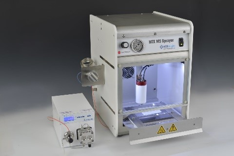
HTX M5 Robotic Reagent Sprayer
The HTX M5 Robotic Reagent Sprayer allows for high-throughput, reproducible sample preparation for MALDI analysis. Reagents such as derivatization agents, proteolytic enzymes, and/or MALDI matrices can be deposited in a uniform manner over tissue sections. Fine control of flow rate, temperature, gas pressure, and nozzle track speed allow for the generation of very small droplets (<5 µm), minimizing delocalization of analytes and allowing for high spatial resolution imaging.

Hamamatsu NanoZoomer SQ
The Hamamatsu NanoZoomer SQ is a high-quality compact digital whole slide scanner. It is capable of scanning a 15x15 mm area at 20x magnification in about 2.5 minutes. It produces seamless brightfield microscopy images that can be directly imported into MSI analysis software. Images can be reviewed and annotated using the NDP.view2 viewer software for region of interest analysis from MSI data.

Thermo NX50 Cryostat
The Thermo NX50 Cryostat allows for high-quality, reproducible, sectioning of frozen tissue specimens. Sectioning is the first step in sample preparation for mass spectrometry imaging and obtaining quality sections is of utmost importance in generating optimal image data. The cryostat allows for section thickness from 0.5 to 100 µm and an operating temperature range from -10 to -43°C. Specimen head alignment minimizes sample loss with repeated sectioning of a block. Adjustable height of the system allows for ergonomic sectioning for all users.
Resources
Sample Submission
The Mass Spectrometry Imaging Facility offers MALDI profiling and imaging of fresh frozen and FFPE tissue sections, as well as profiling of biofluids, cell lines, and liquid biopsies related to the study. It operates under Biosafety Level 2 (BSL2) guidelines, and all submitted samples must comply with BSL2 protocols.
A project begins with a consultation to discuss study goals, optimal instruments, biomolecules of interest, spatial resolution and sample preparation. Early involvement of Facility Personnel is recommended to ensure quality data and determine study scope and cost.
Samples must be dropped off by appointment at Facility (FNT B.101) and given directly to staff for proper storage. Human samples should be de-identified, labeled with a unique code, and transported accordingly (frozen samples on dry ice, FFPE at room temperature). A sample submission form and list (with identifiers, species, tissue type and group info) must be submitted.
Frozen tissues should be submitted as blocks unless prior arrangements for sections have been made. FFPE tissue can be submitted as blocks or sections, with special slide instructions provided if sections are required. Remaining samples will be returned to the investigator at study completion.
Download Sample Submission Form
Guides & Videos
Facility Publications
- Combining Chemistry and Engineering for Hepatocellular Carcinoma: Nano-Scale and Smaller Therapies. Stolley DL, Crouch AC, Ozkan A, Seeley EH, Whitley EM, Rylander MN, Cressman ENK. Pharmaceutics. 2020, 12, 1243.
- Perspective on Mass Spectrometry Imaging for In Situ Visualization of Chemotherapeutics Delivered via Nanoparticle Encapsulation. Seeley EH. Clin Chem. 2021, 67, 1172-1174.
- Reduced Hemoglobin Signal and Improved Detection of Endogenous Proteins in Blood-Rich Tissues for MALDI Mass Spectrometry Imaging. Lin M, Eberlin LS, Seeley EH. J Am Soc Mass Spectrom. 2022, 33, 296-303.
- Are Mass Spectrometry Imaging-Based Diagnostics Becoming Reality? Seeley EH. Proteomics. Clin Appl. 2022, Apr 8:e2200017. doi: 10.1002/prca.202200017.
- Lactate is an Epigenetic Metabolite that Drives Survival in Model Systems of Glioblastoma. Torrini C, Nguyen TTT, Shu C, Mela A, Humala N, Mahajan A, Seeley EH, Zhang G, Westhoff M-A, Karpel-Massler G, Bruce JN, Canoll P, Sieflein MD. Mol Cell. 2022, 82(16), 3061-3076.
- In Situ Imaging of O-Linked B-N-Acetylglucoseamine Using On-Tissue Hydrolysis and MALDI Mass Spectrometry. Escobar EE, Seeley EH, Serrano-Negron JE, Vocadlo DJ, Brodbelt JS. Cancers. 2023, 15(4), 1224-1239.
- Spatially Resolved Metabolites in Stable and Unstable Human Atherosclerotic Plaques Indentified by Mass Spectrometry Imaging. Seeley EH, Liu Z, Yuan S, Stroope C, Cockerham E, Rashdan NA, Delgadillo LF, Finney AC, Kumar D, Das S, Razani B, Liu W, Traylor J, Orr AW, Rom O, Pattillo CB, Yurdagul A Jr. ArteriosclerThromb Vasc Biol. 2023, 43(9), 1626-1635.
- TNFR1 Signaling Converging on FGF14 Controls Neuronal Hyperactivity and Sickness Behavior in Experimental Cerebral Malaria. Dvorak NM, Domingo ND, Tapia CM, Wadsworth PA, Marosi M, Avchalumov Y, Fongsaran C, Koff L, Di Re J, Sampson CM, Baumgartner TJ, Wang P, Villarreal PP, Solomon OD, Stutz SJ, Aditi, Porter J, Gbedande K, Prideaux B, Green TA, Seeley EH, Samir P, Dineley KT, Vargas G, Zhou J, Cisneros I, Stephens R, Laezza F. J Neroinflammation. 2023, 20(1), 306-339.
- Molecular, Metabolic, and Subcellular Mapping of the Tumor Immune Microenvironment via 3D Targeted and Non-Targeted Multiplex Multi-omics Analyeses. Ferri-Borgogno S, Burks JK, Seeley EH, McKee TD, Stolley DL, Basi AV, Gomez JA, Gamal BT, Ayyadhury S, Lawson BC, Yates MS, Birrer MJ, Lu KH, Moc SC. Cancers. 2024, 16(5), 846-868.
- In Vitro Evaluation of the Percutaneous Absorption of Progesterone in Anhydrous Permeation-Enhancing Base using the Franz Skin Finite Does Model and Mass Spectrometry. Banov D, Song G, Ip K, Seeley EH, Linehan ST, Bassani I, Ferron G, Bassani AS, Valdez BC. Arch. Dermatol Res. 2024, 316, 291.
- Spatial Multiplexing and Omics. Carsten JL, Krishnan SN, Rao A, Sorace AG, Seeley EH, Ferri-Borgogno S, Burks JK. Nat Rev Methods Primers. 2024, 4, 54.
- Evaluating Drug Distribution in Rat Lung by Mass Spectrometry Imaging after Dry Powder Insufflation: Comparison of Jet-Milling, Spray-Drying, and Thin Film Freezing. Moon C, Maier EY, Seeley EH, Warnken ZN, Behrend-Keim B, Yanwinitchai S, Mota M, Williams III RO. J Drug Deliv Sci Technol. 2024, 101(A), 106206.
- Inhibition of Hepatic Oxalate Overproduction Ameliorates Metabolic Dysfunction-Associated Steatohepatitis. Das S, Finney AC, Anand SK, Rohilla S, Liu Y, Pandey N, Ghrayeb A, Kumar D, Nunez, K, Liu Z, Arias F, Zhao Y, Pearson-Gallion BH, McKinney MP, Richard KSE, Gomez-Vidal JA, Abdulla CS, Cockerham ED, Eniafe J, Yurochko AD, Tarek Magdy, Pattillo CB, Kevil CG, Razani B, Bhuiyan MS, Seeley EH, Galliano GE, Wei B, Tan L, Mahmud I, Surakka I, Garcia-Barrio MT, Lorenzi PL, Gottlieb E, Salido E, Zhang J, Orr AW, Liu W, Diaz-Gavilan M, Chen YE, Dhanesha N, Thevenot PT, Cohen AJ, Yurdigal Jr A, Rom O. Nat Metab. 2024, 6, 1932-1962.
- Comformational Characterization of Peptides and Proteins by 193 nm Ultraviolet Photodissociation in the Collision Cell of a Trapped Ion Mobility Spectrometry-Time-of-Flight Mass Spectrometer. Butalewicz JP, Escobar EE, Wootton CA, Theisen A, Park MA, Seeley EH, Brodbelt JS. Anal Chem. 2024, 96(41), 16154-16161.
- Environmental exposure to common pesticide induces synaptic deficit and social memory impairment driven by neurodevelopmental vulnerability of hippocampal parvalbumin interneurons. Di Re J, Koff L, Avchalumov Y, Singh AK, Baumgartner TJ, Marosi M, Matz LM, Hallberg LM, Ameredes BT, Seeley EH, Buffington SA, Green TA, Laezza F. J Hazard Mater. 2025, 485, 136893.
- Reevaluating Anti-Inflammatory Therapy: Targeting Senescence to Balance Anti-Cancer Efficacy and Vascular Disease. Casso-Chapa B, Vazquez González NA, Le NT, Palaskas NL, Nead KT, Eutsey LP, Samanthapudi VSK, Osborn AM, Lee J, Mejia G, Hoang O, Lin SH, Deswal A, Herrmann J, Wang G, Kirkland JL, Krishnan S, Wehrens XHT, Chini EN, Yusuf SW, Iliescu CA, Jain A, Burks JK, Seeley E, Lorenzi PL, Chau KM, Ostos-Mendoza KC, Grumbach IM, Brookes PS, Hanssen NMJ, de Winther MPJ, Yvan-Charvet L, Kotla S, Schadler K, Abe JI. Arterioscler Thromb Vasc Biol. 2025, 45(3), 372-385.
- Ketomimetic nutrients remodel the glycocalyx and trigger a metabolic defense in breast cancer cells. Kamra M, Chen YI, Delgado PC, Seeley EH, Seidlits SK, Yeh HC, Brock A, Parekh SH. Cancer Metab. 2025, 13(1), 18.
- Maximizing Data Coverage through Eight Sequential Mass Spectrometry Images of a Single Tissue Section. Seeley EH. J Am Soc Mass Spectrom. 2025, 36(5), 1148-1157.
- Mass spectrometry imaging reveals alterations in protein and N-glycan molecular signatures in endometriosis tissues. Lin M, Seeley EH, Ledet S, Salazar C, Breen MT, Eberlin LS. Anal Bioanal Chem. 2025, Jun 18. doi: 10.1007/s00216-025-05930-z.
Presentations and Posters
- Reduced Hemoglobin Signal and Improved Detection of Low Abundance Proteins in Blood-Rich Tissues for MALDI Mass Spectrometry Imaging. Lin M, Eberlin LS, and Seeley EH. Presented at the 2020 IMSI Poster Gala. December 15-17, 2020. Virtual.
- Improved Diagnosis of Melanocytic Lesions using Mass Spectrometry Imaging. Seeley EH. Presented as part of the HTXNext Webinar Series. June 9, 2021.
- Mass Spectrometry Imaging Enables Localization of Immuno- and Targeted Therapies Delivered Transarterially for Hepatocellular Carcinoma. Seeley EH, Munoz NM, Avritscher R, Koppolu B, Ramkumar N, Johnson R, Kangas S, Sheth RA. Presented at 69th ASMS Conference on Mass Spectrometry and Allied Topics. November 2021. Philadelphia, PA.
- New Sample Preparation Strategies for MALDI Imaging of Clinical Samples. Seeley EH. Bruker/Technology Networks Webinar. December 9, 2021.
- Metabolic Imaging of O-linked N-Acetylglucosamine using On-Tissue Hydrolysis and MALDI. Escobar EE, Seeley EH, Brodbelt JS. Presented at 70th ASMS Conference on Mass Spectrometry and Allied Topics. June 2022. Minneapolis, MN.
- Mass Spectrometry Imaging for Protein Localization and Characterization in Thermoembolized Hepatic Porcine Tissue. Porter JE, Stolley D, Cressman ENK, Seeley EH. Presented at 70th ASMS Conference on Mass Spectrometry and Allied Topics. June 2022. Minneapolis, MN.
- Optimization and Investigation of Protein Molecular Signatures in Endometriosis Tissues by Mass Spectrometry Imaging. Lin M, Seeley EH, Ledet S, Salazar C, Breen MT, Eberlin LS. Presented at 70th ASMS Conference on Mass Spectrometry and Allied Topics. June 2022. Minneapolis, MN.
- Maximizing Data Coverage with Sequential Imaging of a Single Tissue Section. Seeley EH, Escobar EE, Brodbelt JS. Presented at 71st ASMS Conference on Mass Spectrometry and Allied Topics. June 2023. Houston, TX.
- Mass Spectrometry Imaging Reveals Molecular Changes Associated with Aging in Pancreatic Cancer. Seeley EH, Min J, Burks JK, Guerrero PA, Maitra A. Presented at CPRIT Innovations IV. October 2023. Galveston, TX.
- Combining Drug and Targeted Protein Imaging to Uncover Molecular Changes Associated with Cancer Treatment. Seeley EH, Kita CA, Yagnik GB, Rothschild KJ, Lim MJ, Sheth RA. Presented at 72nd ASMS Conference on Mass Spectrometry And Allied Topics. June 2024. Anaheim, CA.
- Fast Lipid Analysis of Lipid A Coupled with Tandem Mass Spectrometry. Zuercher KS, Seeley EH, Brodbelt JS. Presented at 72nd ASMS Conference on Mass Spectrometry And Allied Topics. June 2024. Anaheim, CA.
- Multi-Site Reproducibility Trial of MALDI-IHC Multiplexed Targeted Protein Imaging using a 33-Organ Tissue Microarray. Shafer CC, Holbrook JH, Kita CA, Yagnik GB, Lim MJ, Rosthchild KJ, Stumpo KA, Seeley EH, Neumann EK, Hummon AB. Presented at 72nd ASMS Conference on Mass Spectrometry And Allied Topics. June 2024. Anaheim, CA.
- Spatial Mapping of Ether Lipids in Tissue via MALDI TIMS Imaging Mass Spectrometry: Characterization of Biomarkers of Peroxisomal Disorders. Campbell MJ, Seeley EH, Brodbelt JS. Presented at 72nd ASMS Conference on Mass Spectrometry And Allied Topics. June 2024. Anaheim, CA.
- Spatial Multi-Omics Investigation of High-Grade Serous Ovarian Cancer Tumor Microenvironment Provides Insight into Minimal Residual Disease and Instinsic Chemoresistance. Seeley E, Gamal B, Patterson NH, Zhang W, Mantas MJQ, Ly A, Verbeeck N, Claesen M, Jazaeri A, Burks J, Mok S, Ferri-Borgogno S. Presented at 72nd ASMS Conference on Mass Spectrometry And Allied Topics. June 2024. Anaheim, CA.
- Sequential Imaging and Collagenase Digest Peptide Identification from a Multi-Organ Tissue Microarray. Seeley EH, Bayshal A, Person MD, Pacheco CD, Ly C, Manshouri T, Yin CC, Veletic I. Presented at 73rd ASMS Conference on Mass Spectrometry and Allied Topics. June 2025. Baltimore, MD.
Contact
Email: msimaging@austin.utexas.edu
Phone: 512-232-8189
Operating Hours: Monday-Friday: 8 am to 4:30 pm, appointments strongly recommended.
